Modified Craniotomy Procedures and VR Tech Enhance Brain Surgery
Introduction
Surgical revascularization is crucial for patients with Moyamoya Disease (MMD) and Internal Carotid Artery Occlusion (ICAO). These procedures help improve blood flow to the brain and reduce the risk of strokes. There are three types of revascularization techniques: direct, indirect, and a combination of both.
A key player in these surgeries is the Middle Meningeal Artery (MMA), which can help supply blood to the brain in several ways:
- Donor artery in certain procedures.
- Developing collateral circulation.
- In rare cases, it may even link directly with the ophthalmic artery.
However, the MMA’s complex path and anatomical variations make it susceptible to injuries during surgeries, which can negatively impact patient outcomes.
Challenges in Protecting MMA
Past studies tried using landmarks like the pterion (a region on the skull) to locate the MMA. However, these methods had limitations and didn't always protect the MMA effectively. Advanced techniques like Indocyanine green imaging and surgical navigation systems are accurate but often costly or impractical.
Our Solution: Virtual Reality and Craniotomy Adjustment
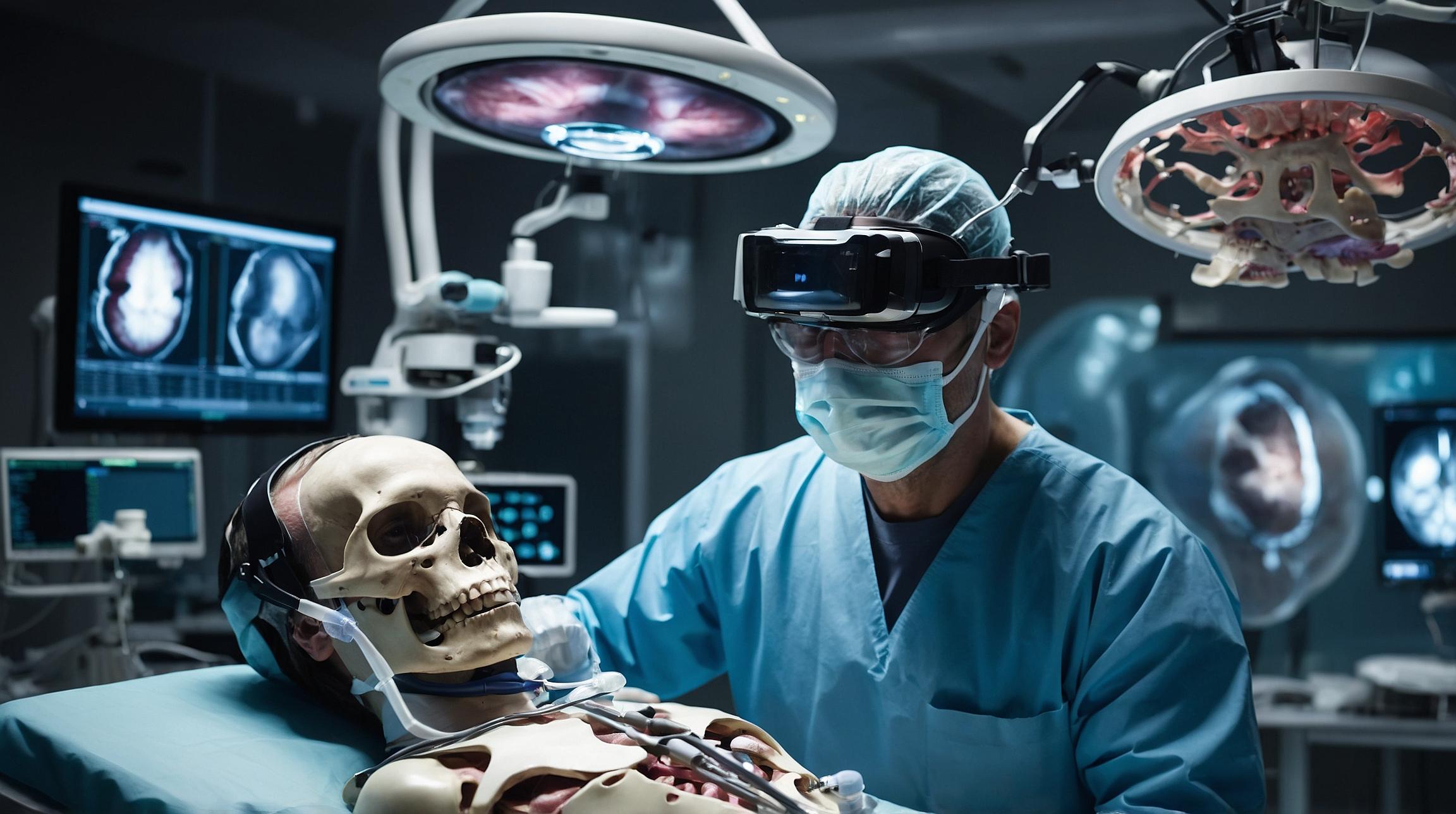
To address these issues, we utilized the free software 3D Slicer along with virtual reality (VR) technology to improve MMA localization. This allows for customized bone flap formation, which can greatly reduce injury risks. This method is cost-effective and widely accessible.
Methods
We studied 71 patients who underwent revascularization between 2015 and 2022. The study had two groups:
- Early group (2015-2020): Used traditional methods.
- Latter group (2020-2022): Employed VR technology for precise MMA localization.
Traditional Methods
The early group relied on landmarks like the Superior Temporal Artery (STA) and coronal suture to estimate the MMA’s location. However, results varied, and the risk of injury remained.
VR-Assisted Methods
In the latter group, we used 3D Slicer software to create 3D skull models from patients' CT scans. Using a mobile app called Digital Brain, we projected these models onto the actual patient’s skull to mark the MMA’s location. This helped us to precisely drill and form bone flaps without damaging the MMA.
Results
Both groups had similar baseline characteristics. However, the MMA preservation rate was significantly higher in the latter group using VR technology (91.7% vs. 68.2% in the early group).
Discussion
Protecting the MMA during surgery is crucial to avoid complications. Our study highlights the importance of accurate localization and suggests that VR technology can be a game changer.
The key takeaways include:
- Increased Preservation: VR-assisted methods significantly improve the preservation rates of the MMA.
- Cost-Effective: This method does not require additional expensive radiological exams.
- User-Friendly: The process is simple and accessible, making it practical for widespread use.
Conclusion
Using modified craniotomy procedures combined with VR technology like 3D Slicer, we achieved more accurate localization and better protection of the MMA in brain surgery. This method is both economical and effective, promising better outcomes for patients with MMD and ICAO.


